 I took my place at the gray table in the gray, poorly-lit room, meeting the gaze of the eyeless skull staked upright before me. A stiff rectangular block of white wax was my only material, and a simple metal implement, sharp like a scalpel on one end and rounded like a spoon on the other, my only tool—other than my own hands and head, once full of vertebrate anatomy knowledge, now less so. To my right, another student, a painter, ripped wax from his block and manipulated it. My weak hands couldn’t get a grip.
Our teacher, Eleanor Crook, an internationally-known fine artist trained in forensic reconstruction, told us to first create the temporalis muscle on one side of the skull, a plaster replica of a real human’s. The muscle is seashell shaped, she explained, fibers radiating upward from near the ear. You can feel this muscle work, if you put your hand on it while opening and closing your jaw.
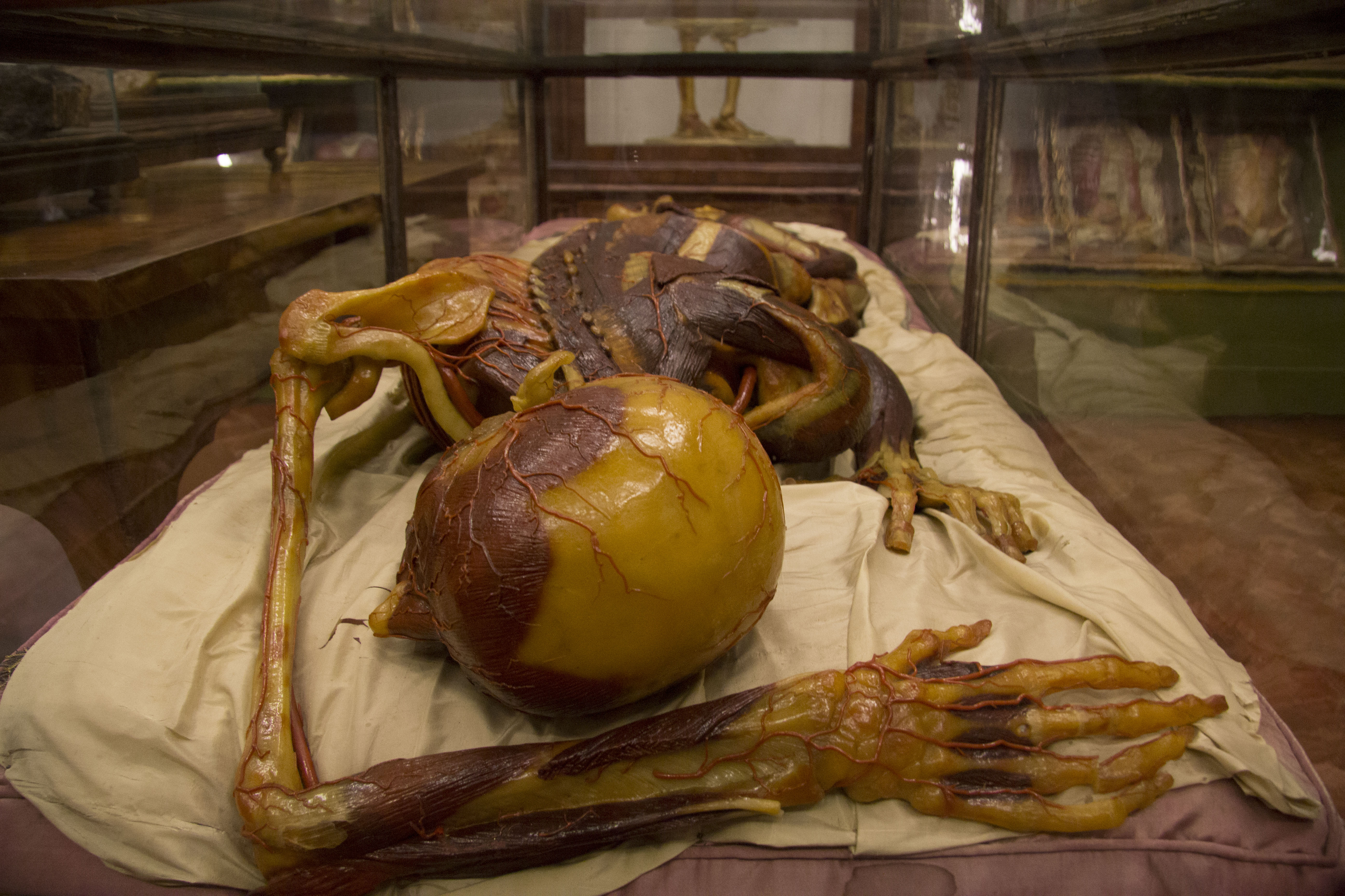
About ten years ago, at a Viennese medical museum, I saw an exhibit of incredibly accurate, life-sized wax anatomical models from the 18th century. That same year, I unexpectedly landed on a modern operating room table, conscious with my innards exposed, and the awful experience sparked an odd feeling of kinship with the models. In my quest to understand their origins, I happened upon this workshop at London’s Hunterian Museum.
But I am not an artist, and I am uncertain how to proceed. I want to create the whole temporalis muscle in my hand, then place it. Eleanor suggests adding a little wax at a time, filling the natural depression in the skull, and sweeping each bit into place. Using the sharp end of the metal tool, I cut a wax chunk from the block, then warm it in my hand, trying to find the optimal malleability between unyielding and too tacky-soft.
Wax is a new material to me, but certainly not to others. People have painted with wax-pigment mixtures for at least 2,000 years and some ancient Egyptian mummy portraits still survive today. In medieval Italy, artists sculpted wax saints and worshippers offered wax votives—often shaped to resemble their ailing body parts—in prayer at churches. During the Enlightenment, Madame Tussaud created lifelike replicas of noblemen and women, and to this day, people make pilgrimages to her commercial shrines to view, and touch, the famous and the infamous. Today, the likenesses of Neanderthals, King Midas, and the shipwrecked victims of Henry VIII’s Mary Rose have all been reconstructed in wax based on skeletal evidence.
I aim to imitate, albeit crudely, the work of the 18th-century Florentine anatomists and artists who made the models I saw in Vienna. Unlike their combination of beeswax, pine resins, scale insect secretions, and pigments, the material in my hands is White #1704, a secret formulation made by British Wax and slightly softer than the one Eleanor had specially made for her exhibitions. Wax is translucent like skin and biddable, she says, her ribbon-wrapped tangle of red hair trailing down her back. She describes the intricacies of facial anatomy, and adds, smiling, You will enjoy making nose cartilages.
But first we move on to the masseter, the part of your cheek that might be called the jowl. I attach bits of wax to the underside of the cheekbone; I bring a lump down to meet the mandible. As each muscle is completed, I score it with the sharp tool to give the illusion of muscle fibers.
I continue to work for two days in this windowless room, molding the head before me, peering occasionally at the surrounding museum cases filled with malformed human skulls atop little skeletons, brains in sparkling jars, and flimsy floating intestines. I’m not sure if the stuffiness and chemical smell is unique to today’s humidity, or if it’s just my unease. We pause to sip tea and munch on cheese and pickled chutney sandwiches.
The bony divots in the skull itself guide our placement of the muscle attachments. But I am less certain when muscles intertwine with one another. I fret about the expression of the lips. I place and replace the eyes in their sockets, unsure. I’m indecisive about how much muscle to conceal with skin, and my first attempt at a scalp resembles a swim cap, so I remove it. Next, I cover the right cheek with skin, and then I clumsily tear a jagged hole with my thumb.
I spent a lot of my education in ecology and evolution taking things apart, seeking understanding. I analyzed specimens pickled, dried, and injected. I enjoyed knowing the static parts, the sounds of the words, and the act of memorizing them…sphenoid, ethmoid, zygomatic arch, and so on. In my dissections of worm, frog, shark, pigeon, cat, cow, I often felt that the tissues themselves showed me where to separate or cut. I could watch the parts isolating themselves. And so I learned.
But there are limits to an approach so singularly focused on dismantling. During my distressing operating-room experience, the surgeon’s hands held centuries of anatomical knowledge as she expertly manipulated my organs and glued my body back together. Yet somehow I was still left in pieces.
At the museum, wax in hand and surrounded by isolated biological specimens, I realize it’s time for me to shift from disassembly to reassembly, something akin to the opposite of dissection. The head before me takes shape, and I fumble toward the whole.
Image: Wikimedia Commons.