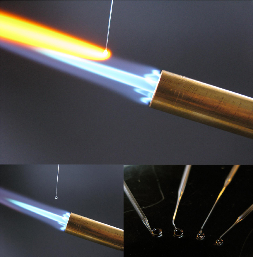
 The University of Victoria conservation field class is rapt. A blowtorch has just been ignited, oomph, and Patrick Keeling, champion of eukaryotes and microbiologist at the University of British Columbia, feeds a straw-thin glass capillary pipette through the hot blue flame. He removes the pipette from the flame and stretches it apart into spun hair. Whoa, the students gasp. Next he snaps the strand of glass in two and feeds one of the ends into the flame again, where its end pools into a little ball “like a lollipop.” An orange comet tail forms in the umbra of the glass. “When your hand gets hot you stop, and that’s your lens,” Keeling says, shaking his hand out and examining his handiwork. He guesses its powers of magnification at 150x. Not shabby.
He breaks off his lens lollipop and sandwiches it between two stamp-sized pieces of paper with pinholes in them. “And just like van Leeuwenhoek did you staple it all together,” he jests (but actually one student buys it. Really? she asks).
Using a piece of sticky tack he mounts a feather onto the back of his DIY microscope and brings the whole apparatus up to his eye. He turns into the sun and his face grimaces in a squint: “Yeah that worked pretty well actually.”
The entire process took about three minutes from start to finish which might make van Leeuwenhoek jump in the nearest lake. Nevertheless, it’s a fitting homage. Leeuwenhoek was an everyday man and Keeling’s lowfi contraption, featured in MAKE magazine and virally spreading across science classrooms the country over, is bringing microscopes not just to eye level, but street level.
 Patrick Keeling, roots for the little guys. Hence why he studies unicellular organisms for a living (and yes, that makes him a protistologist). Cilia, Giardia, flagellum, amoeba, they all make his heart rise in his chest, you can just tell.
The whole reason that microscopic creatures are overlooked so much is that they’re just too small to see. It’s an injustice best remedied by Antonie van Leeuwenhoek, a Dutch bewigged draper who built some 500 single lens microscopes starting in 1673. Therein, Leeuwenhoek found a veritable rabbit hole through which his astounded eyes fell upon a jungle of otherworldly microbes. Leeuwenhoek’s microscopes magnified by 200x, and had fewer aberrations than the compound microscopes that were around at the time.
“I wanted to make a replica of a van Leeuwenhoek microscope for my 3rd year Protistology,” explains Keeling. “Then I realized you could make them out of paper.” Better to make a microscope of your own than look through one already built, he figured.
Around him, the class is abuzz as the students have a go themselves. Blowtorches fire up. Pipettes are broken off. Students root around the grounds of the Hakai Beach Institute, where the class is hosted on Calvert Island in BC’s central coast, looking for odds and end to magnify. Others face the sun like sunflowers, their microscopes pinched in their eye sockets like monocles.
“I like it because I had a lot of boring microscopy classes that I didn’t like,” Keeling says. “You want to get people interested in it but it’s so static.”

In that sense, Keeling has left the world a better place than he found it. Between the blowtorches, the orange-hot glass, and being able to see things at 110x magnification (confirmed by Keeling’s laser pointer) using something you just fashioned out of not household per se but lab-hold items, the class is riveted.
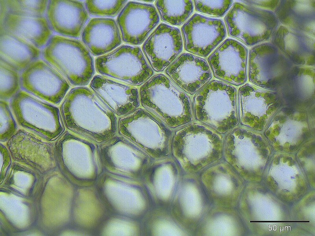
One student finally jiggles her piece of moss into focus. “Wow that is so cool!” she exclaims under her breath to no one in particular. Maybe it’s directed to the world, maybe the tiny cholorophyl-stacked cells that are transformed millimetres from her eyeball, who’s to say.
Not the same words uttered by our Dutch draper perhaps. The sentiment however, that much is unchanged, even 338 years on.
________
Anne Casselman is a Vancouver-based science writer. She writes for various outlets including Canadian Geographic, ScientificAmerican.com, National Geographic News and The Walrus.
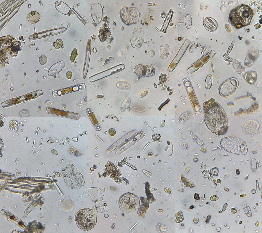
Photo credits: DIY microscope – with kind permission from Patrick Keeling and Erick James; stuff found in a clump of moss – Yersinia; moss cells with chlorophyll – fabelfroh
It is amazing how beautiful the microscopic world is. This is something that students do not always realize when working with their microscopes for the first time. Great images, thanks for sharing!